Sub track:-
Enhanced Image Quality Quantitative Analysis, Faster Turnaround Times,...
Sub track:-
Integration of Imaging Modalities, Advanced Image...
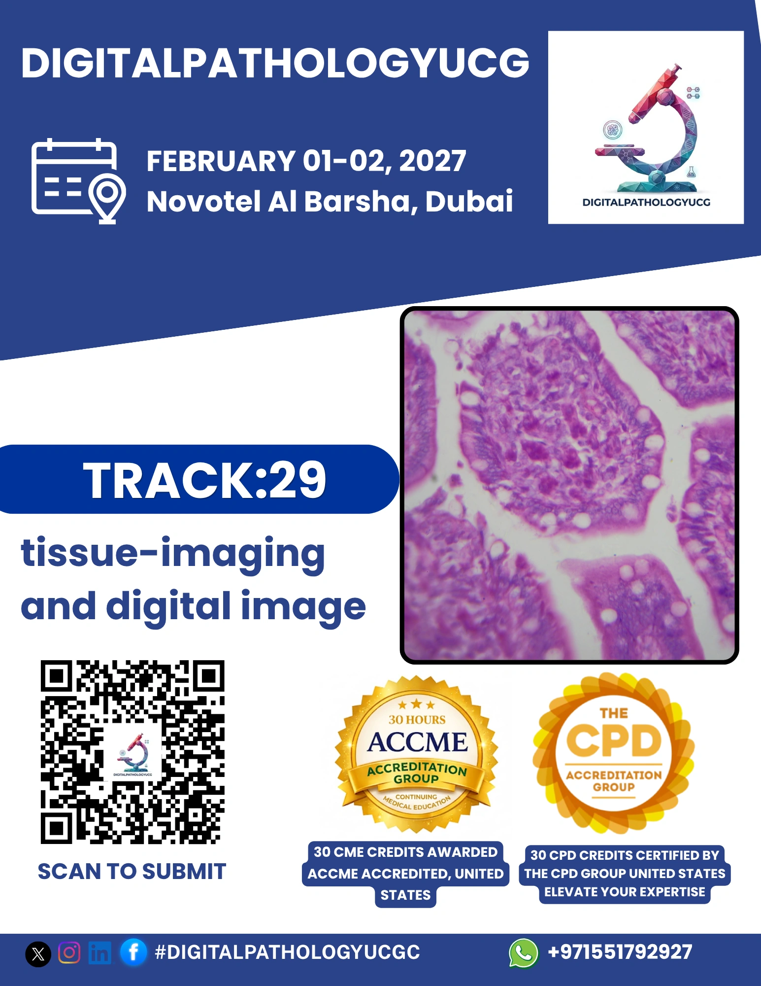
Track Overview:
Tissue imaging and digital image analysis are
fundamental to the advancement of digital pathology. This track focuses on
innovative imaging technologies and computational tools that enhance tissue
visualization, support accurate diagnosis, and accelerate research. From
high-resolution whole slide imaging (WSI) to multiplexed imaging and 3D
reconstruction, digital imaging is transforming how pathologists view,
interpret, and share tissue data.
Key Topics:
Whole slide imaging (WSI) and virtual microscopy
Multiplex immunohistochemistry and
immunofluorescence
3D tissue imaging and volumetric reconstruction
AI-based digital image analysis
Image standardization, annotation, and
interoperability
Data storage, compression, and remote sharing
Learning Objectives:
Understand the latest advancements in tissue
imaging modalities
Explore digital tools that enhance image
interpretation and reporting
Learn how AI supports pattern recognition and
automated quantification
Examine challenges in image management, storage,
and integration
Target Audience:
Pathologists, digital imaging specialists,
researchers, AI developers, lab managers, and professionals in digital
pathology and biomedical imaging.