Sub track:-
Enhanced Image Quality Quantitative Analysis, Faster Turnaround Times,...
Sub track:-
Integration of Imaging Modalities, Advanced Image...
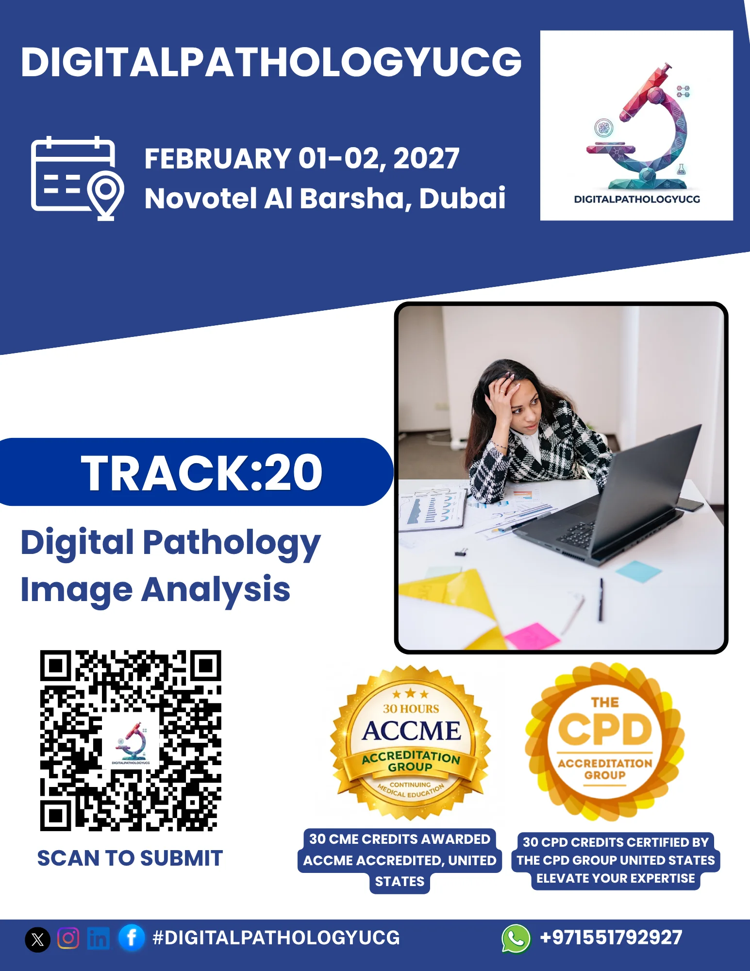
Track Overview:
Digital pathology image analysis is revolutionizing
the way pathologists interpret tissue samples, offering automated,
high-throughput, and precise solutions for evaluating slides. With the
integration of artificial intelligence (AI) and machine learning (ML), digital
image analysis can enhance the detection of abnormalities, improve diagnostic
accuracy, and streamline workflows in pathology labs. This track will explore
the applications of image analysis technologies in digital pathology, focusing
on AI-driven tools, image segmentation, quantification, and their role in
improving patient outcomes.
Key Topics:
Overview of Digital Pathology Image Analysis:
Introduction to the fundamentals of digital pathology image analysis, including
techniques such as image segmentation, feature extraction, and pattern
recognition.
AI and Machine Learning in Image Analysis: How AI
algorithms are transforming digital pathology by automating image
interpretation, detecting patterns, and enhancing diagnostic workflows.
Image Quantification and Biomarker Analysis: Using
image analysis tools for the quantification of tissue structures, tumor cells,
and biomarkers to support diagnosis and treatment decisions.
Applications in Oncology and Other Specialties:
Real-world examples of how image analysis is being applied in cancer
diagnostics, including tumor detection, grading, and prognostication.
Integration with Pathology Workflows: How image
analysis software integrates with digital pathology scanners and laboratory
information systems (LIS) to enhance clinical workflows.
Challenges and Limitations: Addressing the
challenges in digital image analysis, such as data quality, algorithm
validation, standardization, and overcoming biases in AI models.
Learning Objectives:
Gain an understanding of the key principles and
techniques involved in digital pathology image analysis, including AI-based
methods and image processing.
Learn how AI and ML tools are being used to enhance
diagnostic accuracy and automate routine tasks in pathology.
Explore the potential of image analysis in
quantifying tissue features and biomarkers for improved diagnostic and
prognostic outcomes.
Understand the integration of digital image
analysis tools into clinical workflows and their impact on pathology lab
operations.
Discuss the challenges associated with image
analysis, including algorithm validation, regulatory requirements, and ensuring
the reliability of AI systems.
Target Audience:
Pathologists, researchers, and clinicians
interested in the use of image analysis tools for diagnostics.
Data scientists and AI specialists working on the
development and implementation of image analysis algorithms in digital
pathology.
Laboratory managers, healthcare administrators, and
technology providers involved in integrating digital pathology and image
analysis tools into clinical practice.
Regulatory professionals focused on the validation
and approval of AI-based diagnostic tools.
Speakers/Presenters:
Experts in AI and machine learning for digital
pathology image analysis.
Pathologists and clinicians who are using or
developing image analysis tools for clinical applications.
Researchers working on the development of new
algorithms for image segmentation, quantification, and classification in
pathology.
Regulatory specialists and policymakers discussing
standards and guidelines for image analysis tools in healthcare.
Conclusion:
This track will provide valuable insights into the transformative role of digital pathology image analysis in clinical diagnostics. Attendees will learn how AI and machine learning are enhancing diagnostic accuracy, improving workflow efficiency, and enabling more precise and personalized treatment strategies. The track will also address the challenges and future directions in this rapidly advancing field.