Sub track:-
Enhanced Image Quality Quantitative Analysis, Faster Turnaround Times,...
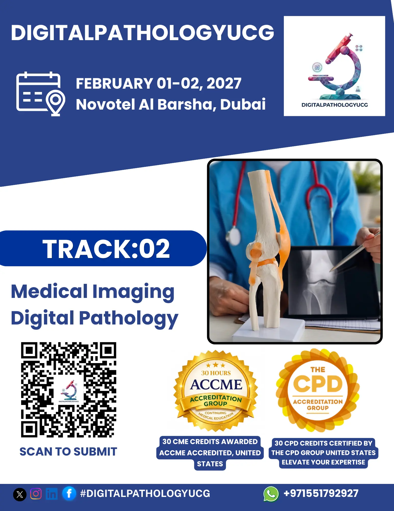
Sub track:-
Integration of Imaging Modalities, Advanced Image...
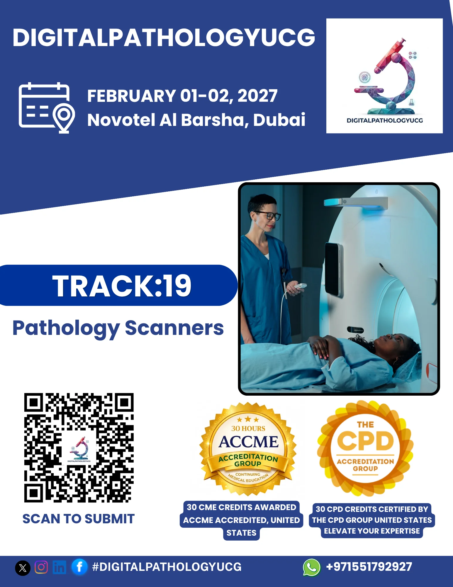
Track Overview:
Pathology scanners, also known as whole-slide
imaging (WSI) systems, are at the heart of digital pathology, enabling
high-resolution, automated scanning of slides for analysis. These devices are
transforming the practice of pathology by digitizing glass slides, facilitating
remote consultations, and enabling the use of advanced image analysis tools.
This track will explore the latest advancements in pathology scanning
technologies, their applications, challenges, and their role in improving
diagnostic accuracy and workflow efficiency in pathology labs.
Key Topics:
Introduction to Pathology Scanners: Overview of the
technology behind pathology scanners, including key features, types of
scanners, and their role in digital pathology workflows.
Technological Advancements in Pathology Scanners:
Exploring the latest developments in scanning resolution, speed, and automation
that have improved the performance and usability of pathology scanners.
Integration with AI and Image Analysis: How
pathology scanners work with AI-driven software to enhance image analysis, such
as automated tumor detection, cell counting, and slide segmentation.
Applications in Clinical and Research Pathology:
Real-world examples of pathology scanners being used in diagnostic practice,
cancer research, and other medical fields.
Challenges in Pathology Scanning: Addressing
challenges such as image quality, scanner calibration, data storage, and
integration with laboratory information systems (LIS).
Regulatory Considerations and Standardization: Understanding
the regulatory landscape for pathology scanners, including FDA approvals,
international standards, and guidelines for clinical use.
Learning Objectives:
Understand the basic principles of pathology
scanners and how they fit into digital pathology workflows.
Learn about the latest innovations in scanning
technology, including advancements in speed, resolution, and AI integration.
Gain insights into how pathology scanners are being
used to improve diagnostic accuracy, particularly in cancer and other complex
diseases.
Discuss the challenges and limitations of pathology
scanning, including image quality, data management, and integration with
existing systems.
Understand the regulatory and standardization
requirements for implementing pathology scanners in clinical practice.
Target Audience:
Pathologists, laboratory managers, and clinical
researchers who use or are interested in using pathology scanners in diagnostic
and research settings.
Technologists and researchers involved in the
development and optimization of scanning equipment and related technologies.
Healthcare administrators and decision-makers
considering the adoption of pathology scanning systems in clinical labs.
Speakers/Presenters:
Experts in pathology scanning technology, including
manufacturers and developers of whole-slide imaging systems.
Pathologists and researchers utilizing pathology
scanners in clinical and research settings.
Regulatory professionals with expertise in the
approval and standardization of digital pathology tools.
AI and machine learning specialists working on
enhancing pathology scanners’ image analysis capabilities.
Conclusion:
This track will provide a comprehensive
understanding of pathology scanners, their technical aspects, applications, and
impact on the pathology field. Attendees will gain practical insights into the
challenges and benefits of using these advanced systems, as well as the future
directions in scanner technology and integration with AI-driven tools.